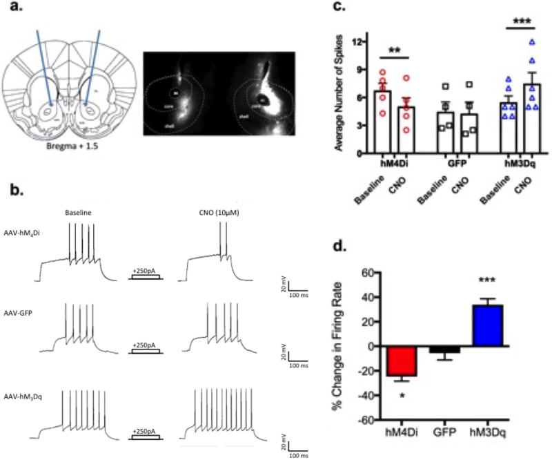
Figure 1. Pharmacogenetic manipulation of neuronal activity in the NAc.
a.) Targeted region for stereotaxic delivery of AAV2 hM3Dq, hM4Di, or GFP to the NAc core+shell (left) and representative 2× image showing viral expression in the NAc core and shell. b.) Representative traces show CNO application reduced the number of evoked action potentials in hM4Di expressing neurons (top), did not change firing rates in GFP expressing neurons (middle), and increased firing in hM3Dq neurons. c.) individual and mean firing rates for each group at baseline and after CNO (n=4–6/group; Two-way ANOVA treatment×group interaction, F(2,12)=22.8, p<0.0001). Bonferroni post-hoc **=p<0.01, ***p<0.001 (CNO vs.baseline). d.) Percent change in firing rate after CNO application (One-way ANOVA F(2,12)=42.8, p<0.0001); Bonferroni post-hoc *=p<0.05 (hM4Di vs GFP), ***p<0.001 (hM3Dq vs. GFP).

