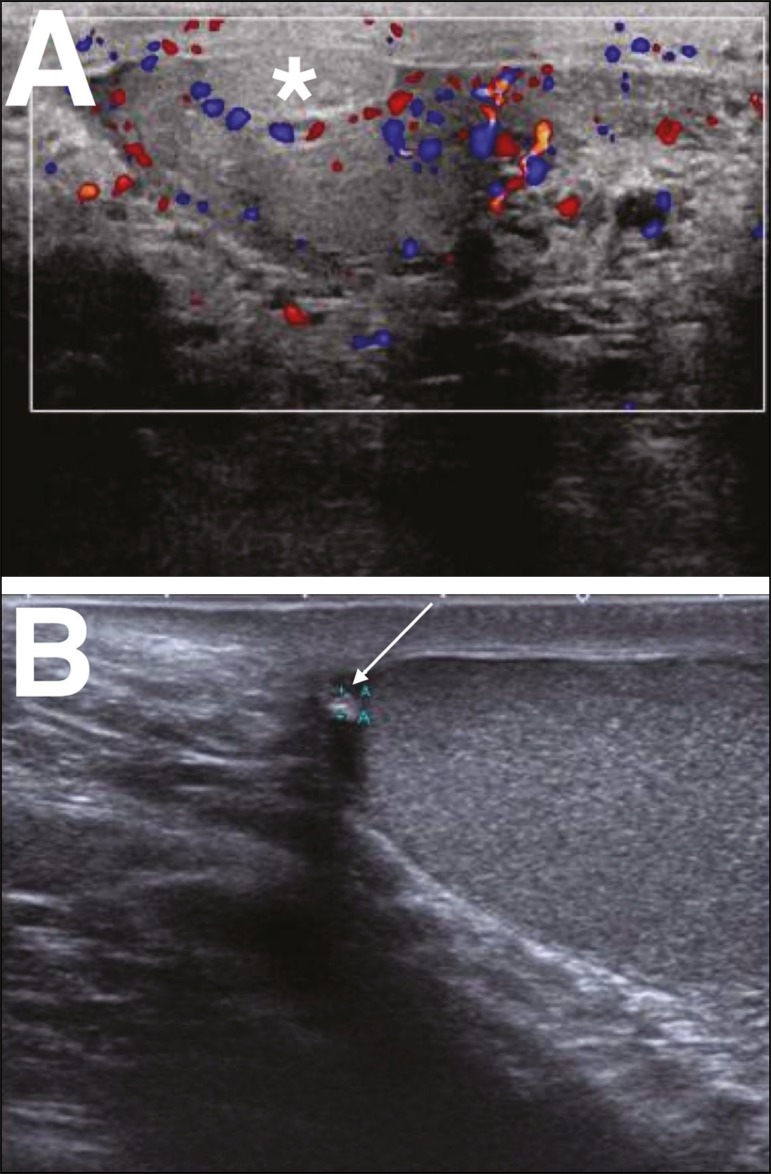
Figure 6.
A: Torsion of the appendix testis. Note the enlargement of the appendix testis (asterisk), showing no flow on the Doppler flow study. The adjacent testis presents only reactive hyperemia, without alteration of its echotexture. B: Chronic torsion of the appendix testis. Note the increased echogenicity and decreased size of the appendix testis (arrow).

