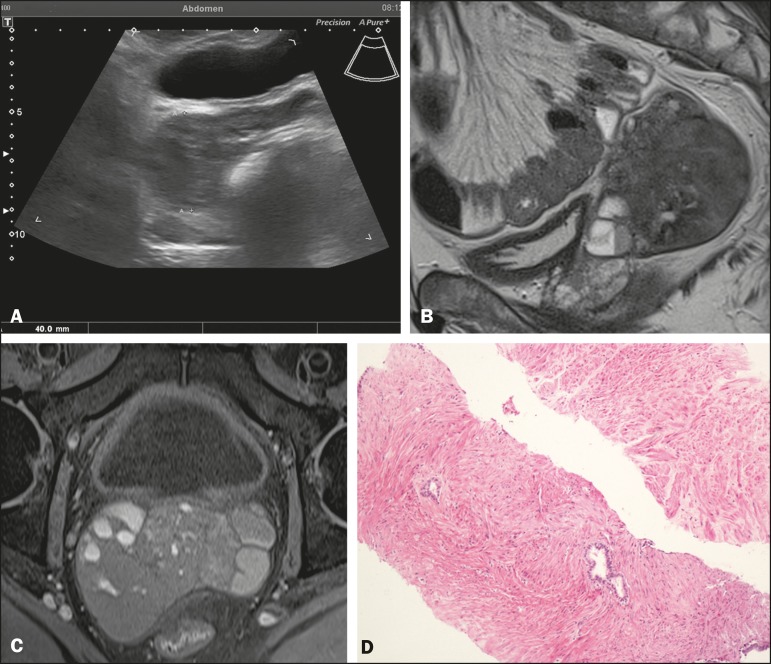
Figure 1.
A: Transabdominal pelvic ultrasound showing a well-defined solid hypoechoic lesion, measuring 4.0 cm, in the right seminal vesicle space. B: Histological slide showing abundant, benign mature smooth muscle, with scanty bistratified columnar epithelium. C: Sagittal T2-weighted MRI sequence showing a well-defined, heterogeneous expansile lesion with predominantly low signal intensity. Note also the fluid-fluid level. D: Axial T1- weighted fast spin-echo MRI sequence showing a solid heterogeneous lesion with its epicenter in the right seminal vesicle and a predominantly isointense signal.

