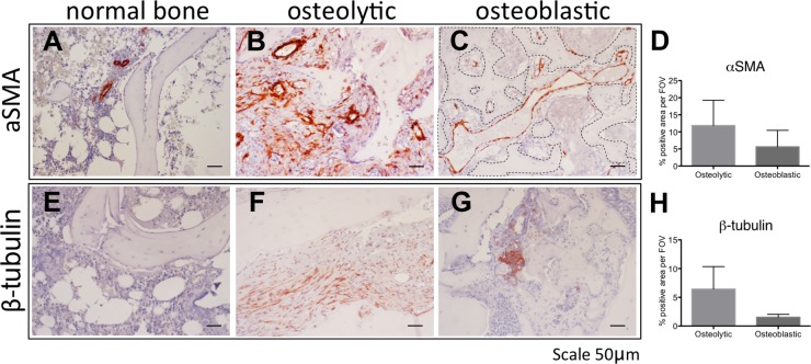
Figure 7. The stroma of human osteolytic bone metastases consist of arteries.
Immunohistochemical detection of αSMA (A-C) and β3 tubulin (E-G) in normal bone (A, E), in osteolytic bone metastasis (B, F) and in osteoblastic bone metastasis (C, G). Scale bar = 50 μm. Positive staining area of αSMA (D) and β3 tubulin (H) was quantified in 2 specimens of osteoblastic bone metastasis and 3 specimens of osteolytic bone metastasis. Dotted line in panel C highlights the bone surface.

