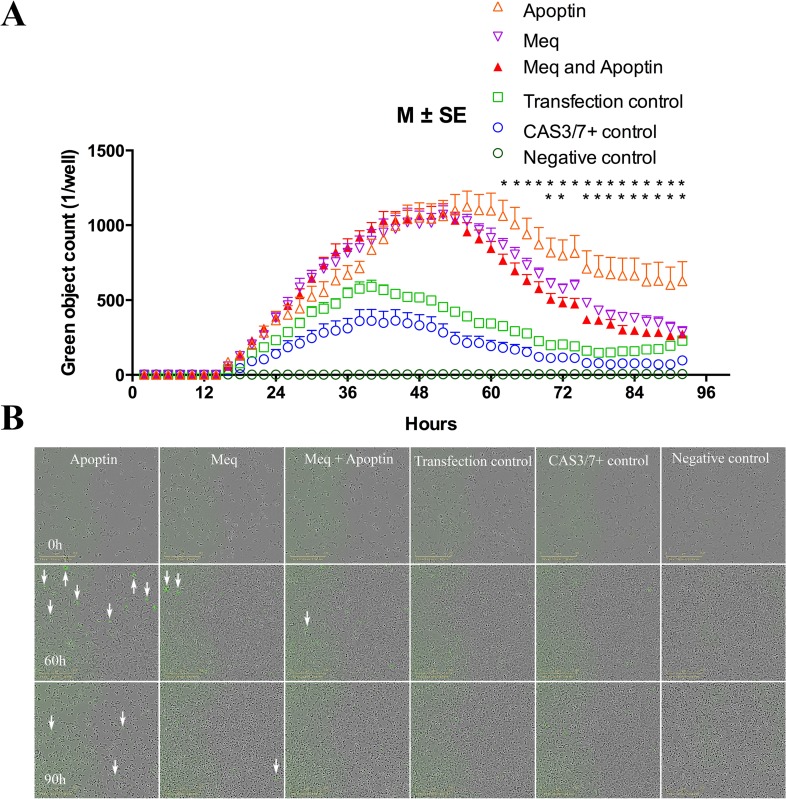
Figure 4. Meq, Apoptin, Meq-Apoptin plasmid transfected DF-1 cells were monitored in real time by the live cells imaging system IncuCyte.
(A) Meq-Apoptin co-transfected cells apoptosis was significantly lower than Apoptin transfected cells between 60 and 92 h. And Meq-transfected cells apoptosis was significantly lower compared to Apoptin-transfected cells between 68 and 92 h, except at 74 h. Growth curves are shown as means of six independent experiments ± standard error (SE). An asterisk (*) indicates statistically significant difference (P < 0.05) between Apoptin and Meq-Apoptin or Meq transfected cells. (B) Representative IncuCyte live cells images illustrating evolution of apoptotic cells (green) at 0, 60 and 90 h. Transfection control, Caspase 3/7 positive control and negative control cells were used as controls.

