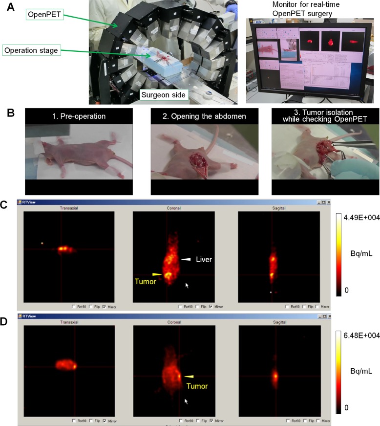
Figure 3. OpenPET-guided surgery.
(A) OpenPET system used for the surgical procedures. (B) Representative images of mice bearing transplanted HCT116-RFP tumors at deep sites during OpenPET-guided surgery. A series of images taken during OpenPET-guided surgery is shown, including pre-operation (left), opening the abdomen (middle), and tumor isolation while checking real-time OpenPET images (right) (see details in Supplementary Video 1). (C, D) OpenPET images. (C) A tumor (approximately 10 mm) located behind the intestine in the middle of peritoneal cavity is shown. (D) A small (3-mm) tumor located deeply in the peritoneal cavity is shown.

