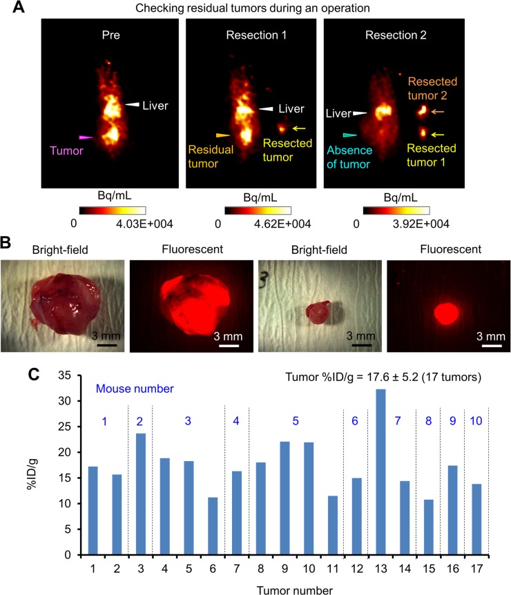
Figure 4. Tumor resection during OpenPET-guided surgery.
(A) A series of OpenPET images taken during tumor resection in OpenPET-guided surgery using mice bearing transplanted HCT116-RFP tumors. These images were obtained before the operation (left), after the first tumor resection (middle), and after the second tumor resection (right). After the first resection, a residual tumor was found by OpenPET. After the second tumor resection, no tumor signals were observed by OpenPET. (B) Bright-field and fluorescence images of resected tumors observed with a stereoscopic fluorescence microscope (10-mm and 3-mm tumors, left and right images). (C) The uptake of 64Cu-PCTA-cetuximab by 17 tumors resected from 10 different mice during OpenPET-guided surgery (%ID/g). The average %ID/g was 17.6 ± 5.2.

