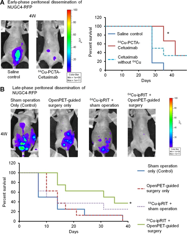
Figure 6. Integrated 64Cu therapy for peritoneal-dissemination models with NUGC4-RFP cells.
(A) The in vivo study of 64Cu-ipRIT with an early-phase peritoneal-dissemination model with NUGC4-RFP cells (saline control, 64Cu-PCTA-cetuximab, cetuximab without 64Cu). Representative images of mice observed by in vivo fluorescence imaging in the saline-control and 64Cu-PCTA-cetuximab groups at 4 weeks after treatment (left). Survival curves (n = 6) (right). *P < 0.05 vs. saline control (log-rank test). (B) The in vivo study of combined treatment with 64Cu-ipRIT and OpenPET-guided surgery with a late-phase peritoneal-dissemination model using NUGC4-RFP cells (sham operation-only control, OpenPET-guided surgery only, 64Cu-ipRIT + sham operation, or 64Cu-ipRIT + OpenPET-guided surgery). Representative images of mice observed by in vivo fluorescence imaging in different groups (sham operation-only control, OpenPET-guided surgery only, 64Cu-ipRIT + sham operation, and 64Cu-ipRIT + OpenPET-guided surgery) at 4 weeks after treatment (upper). Survival curves (n = 8) (lower). *P < 0.05 vs. sham operation-only control (log-rank test)

