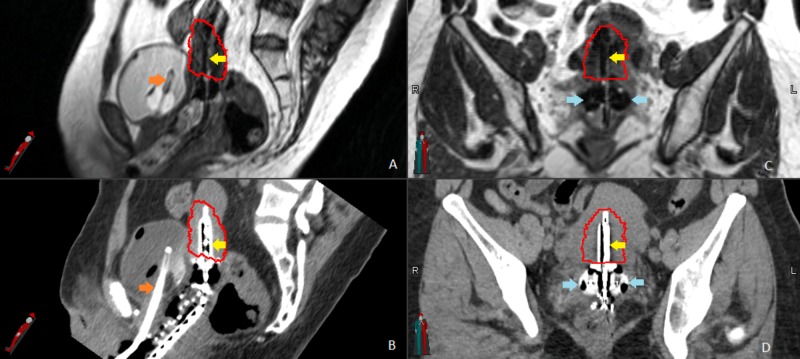
Figure 1. Magnetic resonance-guided brachytherapy.
(A) and (B) show the uterine tandem (yellow arrows) and foley catheter (orange arrows) in the sagittal plan on the magnetic resonance imaging (MRI) and computed tomography (CT). The high-risk clinical target volume (HR-CTV) is outlined in red. (C) and (D) show HR-CTV, tandem (yellow arrows), and ovoids (blue arrows) in the coronal plane, again on the respective MRI and CT.

