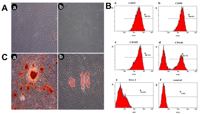
Fig 1. Evaluation of stem cell phenotype.
A (Aa) Third-generation cells(x4), (Ab) Third-generation cells (x10); B Detection of cell surface marker characteristic of mesenchymal stem cells on PDLSC Analyses were performed via flow cytometry detecting FITC conjugated monoclonal antibodies for human CD29, CD90, CD105, CD146, Stro-1(Ba, Bb, Bc, Bd, Be), and negative control(Bf); C (Ca) the formation of calcified nodules, (Cb) The formation of lipid droplets.

