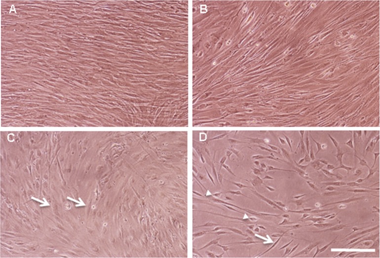
Fig 2. Cultures of hFIBs.

Photomicrographs of living hFIB cultures grown in hFIB Med (A and B) or in RA&EGF-containing SFM (C and D) for two weeks. (A and C) Non-infected control cultures; (B and D) GPA hFIBs transduced with lentiviral particles carrying the GFI1, Pou4f3 and ATOH1 ORFs. Non-infected control cultures (A) and GPA transduced hFIBs (B) grown in hFIB Med showed densely packed fusiform cells, while in the presence of RA&EGF-containing SFM (C) a lower cell density was observed with cells that appeared more flattened out (arrows). (D) GPA transduced hFIB cultures grown in RA&EGF-containing SFM showed a further reduction in cell density and the presence of birefringent elongated (arrow) and smaller polygonal cells (arrowheads). Scale bar: 70μm.
