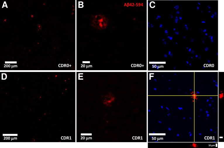
Fig 2. Exemplar fluorescent confocal microscopy of Aβ1-42-biotin binding to unfixed, frozen frontal cortex sections.
(A, D) Representative fluorescent confocal images of labelled (streptavidin-Alexa594, red channel) Aβ1-42-biotin binding in elderly high-pathology controls (CDR0 + plaques) and elderly subjects with mild ADD (CDR1). Scale bar = 200 μm. (B, E) Higher magnification confocal images reveal distinct plaque morphology and minimal background fluorescence in both in elderly high-pathology controls (CDR0 + plaques) and elderly subjects with mild ADD (CDR1). Scale bar = 20 μm. (C) Fluorescent images of labelled (streptavidin-Alexa594, red channel) Aβ1-42-biotin binding display an absence of plaque structure morphology with minimal background signal in cognitively normal elderly subjects without plaque pathology. Nuclei stained with DAPI (blue channel). Scale bar = 50 μm. (F) Fluorescent images of labelled (streptavidin-Alexa594, red channel) Aβ1-42-biotin binding display punctate staining of a central core with peripheral decoration in elderly subjects with mild ADD (CDR1). Nuclei stained with DAPI (blue channel). Scale bar = 50 μm. Orthogonal XZ and YZ views, centered on the yellow crosshairs, demonstrate the labelling extent through the tissue section. Scale bar = 10 μm.

