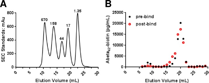
Fig 5. Size exclusion chromatography reveals that the Aβ1-42-biotin monomer remained predominantly low molecular weight during the experiment.
(A) Globular protein molecular weight standards as run on a size exclusion Superdex 200 10/300 GL column. mAu = milli-absorbance units, kDa = kilodaltons. (B) The predominance of the pre-incubation (black closed circles) Aβ1-42-biotin elutes as a low molecular weight peak in fractions 18 to 21 likely corresponding to monomer, and a minority of the Aβ1-42-biotin eluted as a high molecular weight peak in fractions 8 and 9 corresponding to aggregated Aβ. The post overnight incubation sample (red open circles) has a minor shift to include a shoulder of fractions 16 and 17 in the low molecular weight peak suggesting the accumulation of oligomeric species while most the sample remained monomeric.

