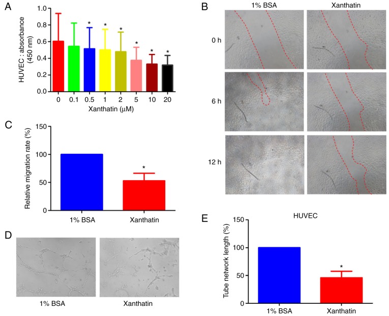
Figure 1.
(A) HUVEC proliferation following exposure to different concentrations of xanthatin for 24 h was detected using a CCK-8 assay. (B) Xanthatin reduced wound closure in a scratch wounding assay of the HUVECs. Images were captured at 0, 6 and 12 h (magnification, ×20) and (C) quantified. (D) Effect of xanthatin on tube formation of HUVECs was assessed by a tubulogenesis assay. Images were captured following treatment with xanthatin for 6 h. (E) Representative images from three independent experiments performed in duplicate. All data are presented as the mean ± standard deviation from three experiments. *P<0.05, compared with the control group. HUVECs, human umbilical vein endothelial cells; BSA, bovine serum albumin.

