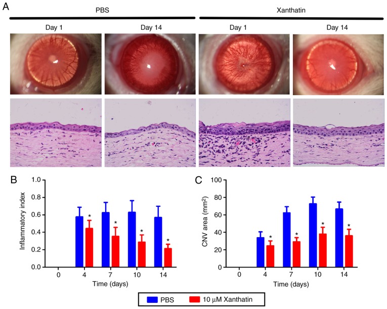
Figure 3.
Inhibition of alkali burn-induced CNV and inflammation by xanthatin treatment. (A) Stereomicroscopic appearance of mouse eyes of the xanthatin group and the control group on days 1 and 14. Representative hematoxylin and eosin-stained corneal sections of the rats in the xanthatin group and the control group are shown. Magnification, ×20. (B) Inflammatory indices of the xanthatin group and the control group were accessed on day 0, 4, 7, 10 and 14. (C) A significant regression of the CNV area was observed in the xanthatin group, compared with that in the PBS treatment on days 0, 4, 7, 10 and 14. All data are presented as the mean ± standard deviation from three experiments. *P<0.05, compared with the control group. CNV, corneal neovascularization; D, day.

