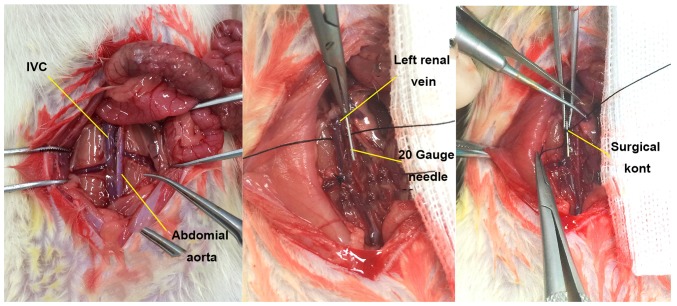
Figure 1.
Anatomy and modeling of IVC in rats. The IVC was disserted and exposed, and a 20-gauge (d=0.91 mm) syringe needle was placed in parallel. The IVC and the needle were ligated with 7-0 polypropylene sutures at ~1 mm below the left renal vein, and then the needle was removed for partial flow restriction of the blood. IVC, inferior vena cava.

