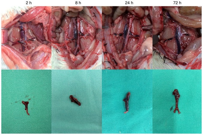
Figure 2.
Thrombosis in the deep venous thrombosis model group. At 2 h, the whole segment of the vein was dark red, with the coagulum in the lumen moving when the lumen wall was compressed. At 8 h, the vein had darkened and the coagulum was harder. At 24 h, the vein appeared black red, the thrombosis inside the vein remained tough, the vein demonstrated decreased mobility and inflammatory manifestation. At 72 h, the vein and embolus were stiff and dark black.

