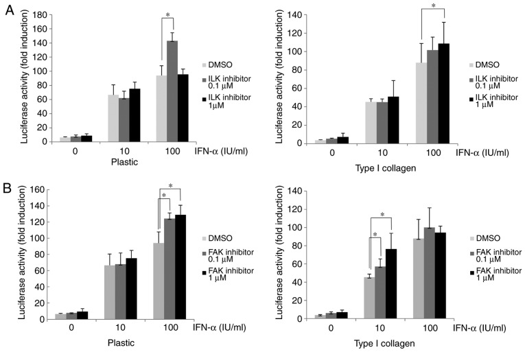
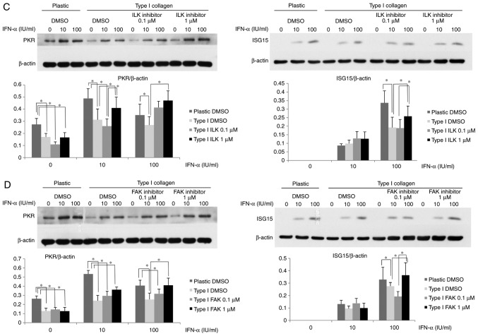
Figure 4.
Attenuation of IFN-α-signaling in HuH-7 cells by type I collagen involves ILK and FAK. (A and B) Improvement in the ISRE-luciferase activity after treatment with (A) ILK inhibitor or (B) FAK inhibitor in HuH-7 cells cultured on normal plastic dishes or type I collagen-coated dishes. Cells were treated with ILK or FAK inhibitor for 6 h. The ISRE-luciferase activity was measured after IFN-α treatment for 12 h. (C and D) Improvement in the ISG protein expression by treatment of (C) ILK inhibitor or (D) FAK inhibitor in OR6 cells grown on normal plastic dishes or type I collagen-coated dishes. OR6 cells were cultured for 3 days and then treated with ILK or FAK inhibitor for 6 h, followed by treatment with IFN-α for 12 h. The expression of ISG15 and PKR was measured by western blot analysis with β-actin used as a control. Values are expressed as the mean ± standard deviation (n=3). *P<0.05. DMSO, dimethyl sulfoxide; ILK, integrin-linked kinase; FAK, focal adhesion kinase; IFN, interferon; OR6 cells, HuH-7 cells stably transfected with full-length HCV-RNA fused with Renilla luciferase ISRE, IFN-stimulated response element; ISG, IFN-stimulated gene; PKR, protein kinase R.


