Figure 1.
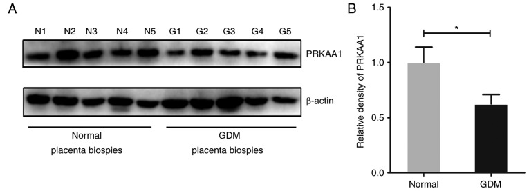
PRKAA1 is decreased in placental tissues of women with GDM. Western blot analysis was used to measure the protein level of PRKAA1 in placental tissues of women without GDM (n=11) and with GDM (n=11). β-actin (~43 kDa) was used as the internal control. The relative density of PRKAA1 (~62 kDa) was determined with Image Pro Plus version 6.0 software. (A) Representative protein bands of PRKAA1 and β-actin in placental biopsies of five normal pregnant women and five women with GDM. (B) Relative density of PRKAA1 in placental tissues of the normal (n=11) and GDM (n=11) groups. Data are expressed as the mean ± standard error of the mean; statistical significance was determined using Student's t-test, *P<0.05. GDM, gestational diabetes mellitus; PRKAA1, protein kinase AMP-activated catalytic subunit α1; N, normal; G, GDM.

