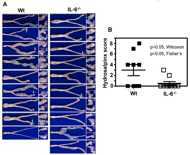
Fig.4. Comparison of hydrosalpinx development between mice with or without IL-6 deficiency following C. muridarum infection at a low dose.

Groups of female C57BL/6J mice without (left column, wild type or Wt, n=9) or with (right column, IL-6−/−,,n=10) deficiency in IL-6 were inoculated intravaginally with 5 × 10³ IFUs of C. muridarum as described in Fig.3 legend. On day 63, all mice were sacrificed for observing upper genital tract pathology hydrosalpinx. (A) Each individual mouse genital tract was shown with the vagina pointing to the left and oviduct/ovary to the right side. Oviduct with hydrosalpinx was indicated with a white arrow and the oviduct/ovary region was magnified shown on the right with the corresponding hydrosalpinx scores indicated. (B) Hydrosalpinx scores (p<0.05, Wilcoxon) and incidences (p>0.05, Fisher’s Exact) were compared between Wt (solid bar) and IL-6−/− (open bar) mice.
