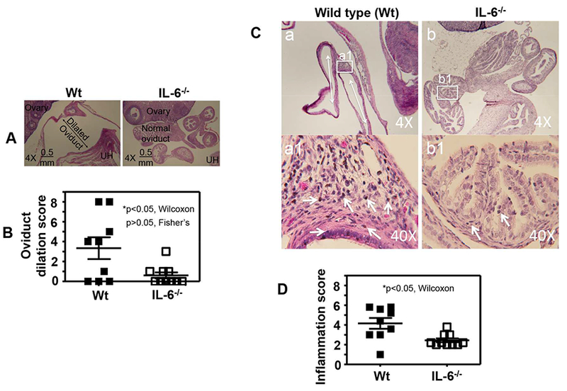
Fig.5. Comparison of oviduct dilation and inflammatory infiltration between mice with or without IL-6 deficiency following C. muridarum infection at a low dose.

Groups of female C57BL/6J mice were inoculated intravaginally with 5 × 10³ IFUs of C. muridarum as described in Fig.4 legend. On day 63, following documenting the macroscopic pathology hydrosalpinx, the same genital tract tissues were used to make sections for microscopic evaluation of oviduct dilation using the criteria described in the materials and methods section. (A) Representative microscopic images of the oviduct area acquired under a 4× objective lens from the mouse groups without (left column, wild type or Wt, n=9) or with (right column, IL-6−/−,,n=10) deficiency in IL-6 were shown with the dilated and normal oviduct, ovary and uterine horn (UH) marked. (B) Oviduct dilation scores (p<0.05, Wilcoxon) and incidences (p>0.05, Fisher’s Exact) were compared between Wt (solid bar) and IL-6−/− (open bar) mice. (C) Representative microscopic images of the oviduct area acquired under 4× or 40× objective lens from the mouse groups without (left column, wild type or Wt, n=9) or with (right column, IL-6−/−,, n=10) deficiency in IL-6 were shown with inflammatory infiltrates marked. The tissue areas that were used to semiquantitate inflammation under 40× were marked in the 4× images. (D) Oviduct inflammatory scores were compared between Wt (solid bar) and IL-6−/− (open bar) mice (*p<0.05, Wilcoxon).
