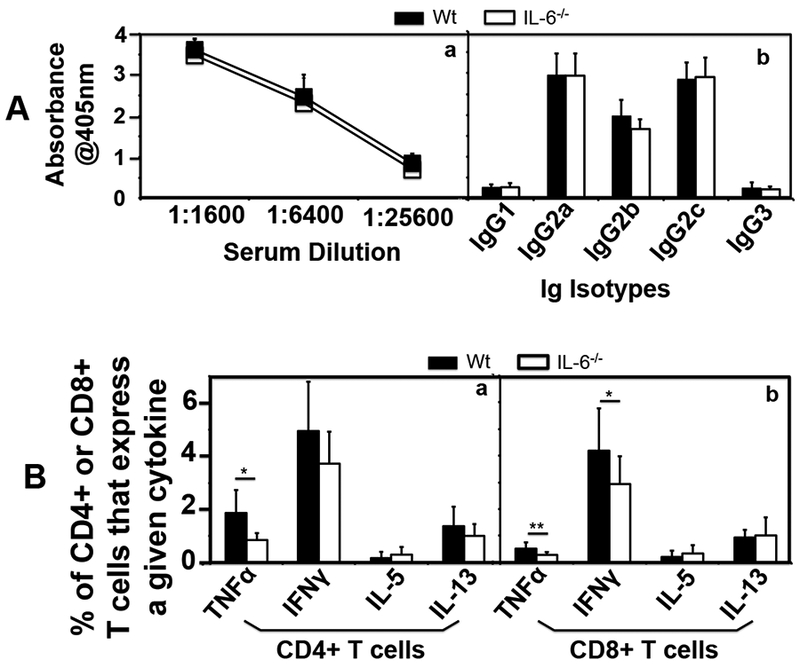
Fig.6. Comparison of anti-C. muridarum antibody production and T cell responses between mice with or without IL-6 deficiency following C. muridarum infection at a low dose.

Groups of female C57BL/6J mice without (solid square or bar, wild type or Wt, n=9) or with (open square or bar IL-6−/−,, n=10) deficiency in IL-6 were inoculated intravaginally with 5 × 10³ IFUs of C. muridarum as described in Fig. 3 legend. (A) On day 63, blood was collected for measuring anti-C. muridarum antibodies in sera using ELISA. The results were expressed as absorbance acquired at the wavelength of 405nm. Total IgG antibodies were measured after the serially diluted serum samples were added to the C. muridarum EB-coated 96 well plates (a) while the serum IgG isotypes were measured using the 1:1600 diluted mouse serum samples and the corresponding mouse IgG isotype-specific HRP conjugates (b). Note that there was no significant difference in the levels of either total IgG or IgG isotypes between Wt and IL-6−/− groups. All mice developed IgG2 isotype-dominant anti-C. muridarum antibody responses. (B) Splenocytes from the same mice were collected for measuring intracellular cytokines (as listed along the X-axis) using flow cytometry from both CD4+ (panel a) and CD8+ (b) T cells following in vitro restimulation with C. muridarum organism-pulsed DCs. The results were expressed as % of CD4+ or CD8+ T cells that produced a given cytokine. Note that wild type mice developed more CD4+ or CD8+ T cells that produce TNFα or IFNγ in response to an in vitro re-stimulation (*p<0.05, **p<0.01). Please note that although the difference in IFNγ-producing CD4+ T cells between the wild type and IL-6-deficient mice was not statistically different, the trend of decrease in IFNγ-producing CD4+ T cells was obvious in the IL-6-deficient mice.
