
Figure 6.
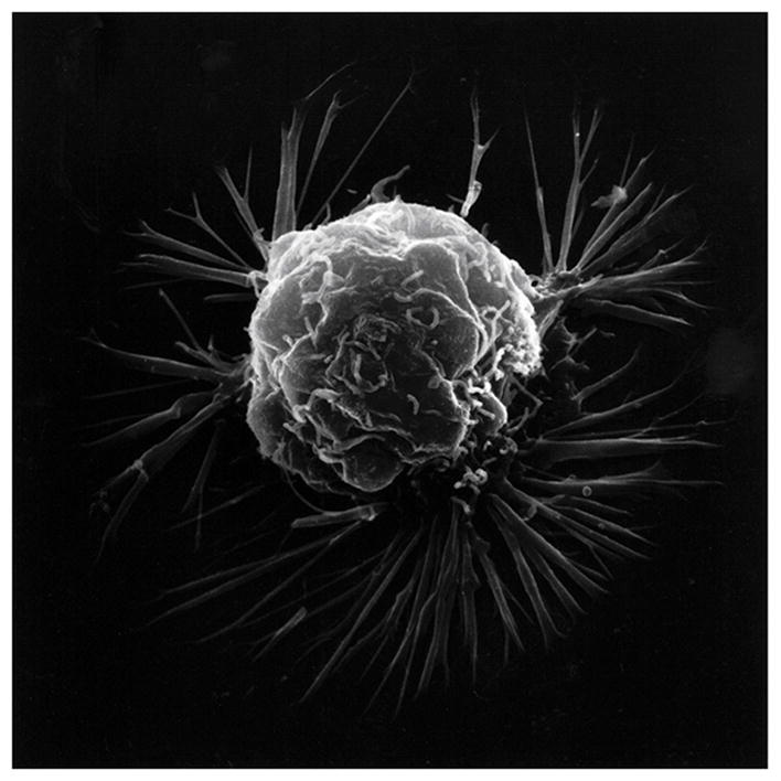
Scanning electron microscopy of an isolated cancer cell with membrane ruffling and long lamellipodia spike-like extensions. (With permission from the National Institutes of Health/Department of Health and Human Services).
Official websites use .gov
A
.gov website belongs to an official
government organization in the United States.
Secure .gov websites use HTTPS
A lock (
) or https:// means you've safely
connected to the .gov website. Share sensitive
information only on official, secure websites.
Scanning electron microscopy of an isolated cancer cell with membrane ruffling and long lamellipodia spike-like extensions. (With permission from the National Institutes of Health/Department of Health and Human Services).