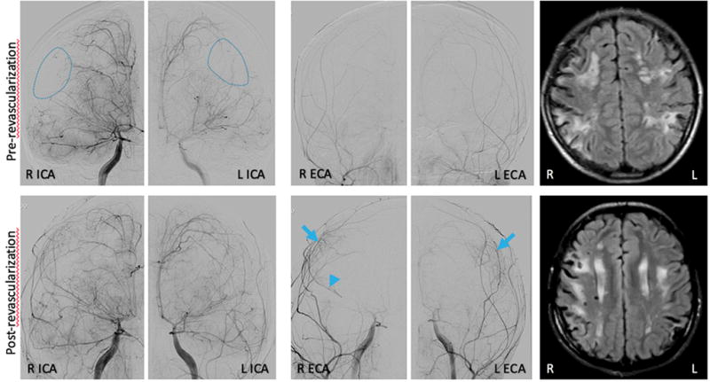
Figure 3.

Representative images before (upper panel) and one year after (low panel) indirect revascularization surgery (dural inversion). Angiographic images show multiple abnormalities including straightening of cerebral arteries, dilation of the petrous and cavernous portions followed by severe stenosis of the terminal segment of the right (R) and left (L) internal carotid arteries (ICA), severe stenosis of the middle and anterior cerebral arteries (with distal R M1 occlusion) and areas of parenchymal hypoperfusion (dashed circles). Follow-up angiogram a year after surgery demonstrates new collateral vessels arising from the middle meningeal arteries (arrows) in the watershed regions of hypoperfusion, with concomitant enlargement of the parent middle meningeal vessel indicating compensatory increased flow, and retrograde flow through the R MCA (arrowhead) supporting hemodynamically significant supply from the donor vessels. MRI FLAIR images illustrate white matter injury and ischemic infarcts at both time points.
