
Figure 9. Cellular localization of HSA-28P.
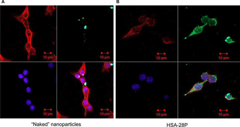
INS-1E cells were treated with FITC-labeled “naked” NP (green) (A) or with FITC-labeled HSA-28P (B) and then fixed as described in “Methods”. The obtained slides were stained with Alexa Fluor 633 Phalloidin for membrane (red) and with DAPI for nuclei (blue), as described in “Methods”. Cells were then visualized with a Confocal-Zeiss microscope. Representative images are shown. n=6.