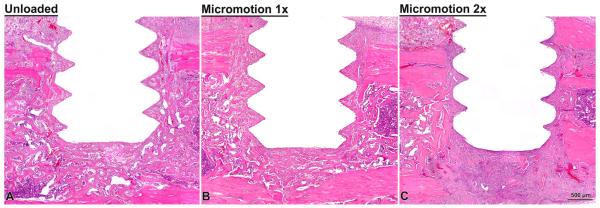
Fig. 3.
Light microscope images of decalcified sections, stained with HE, from the unloaded (A), micromotion 1× (B) and micromotion 2× (C) groups at 7 days post-surgery. Histological observations revealed that new bone forms around the implant in all groups, including between the implants threads. However, signs of disruption of bone healing at the bone/implant interface were noticed in all the animals from the Micromotion 2× group.

