
Figure 2. Radiation induces proliferation and villus differentiation in a subset of Paneth cells.
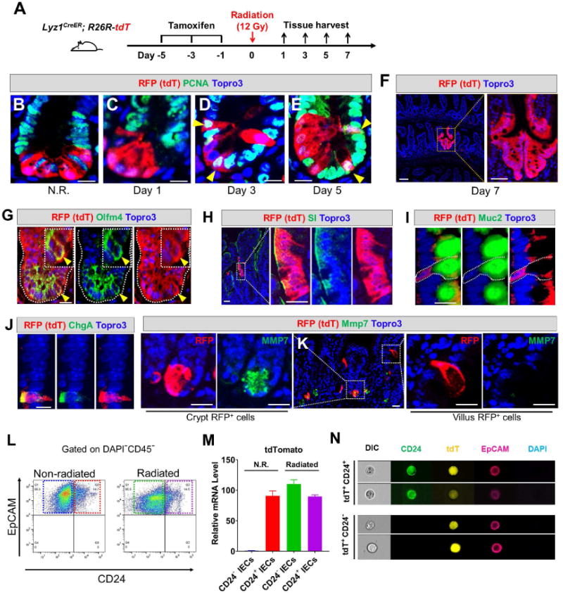
(A) Experimental scheme showing lineage labeling of adult Lyz1CreER; R26R-tdT mice by tamoxifen injection, followed by radiation and tissue analyses (3 mice for each time point).
(B-E) Non-irradiated (N.R.) tdT+ Paneth cells do not express PCNA, however after irradiation tdT+ cells became positive for PCNA on day 3 and 5 (yellow arrowheads). Scale bar, 10 μm.
(F) Radiated Lyz1CreER; R26R-tdT mice showed red epithelial stripes (a representative image of day 7 was shown).
(G-J) Some irradiated tdT+ Paneth cells co-expressed Olfm4 (yellow arrowheads in G) in the crypts, while some villus tdT+ cells co-expressed SI (H), or Muc2 (I). or ChgA (J). Scale bar, 10 μm.
(K) Some irradiated tdT+ cells retained Paneth cell marker (Mmp7, left panel), while villus tdT+ cells lost Paneth cell marker (right panel). Scale bar, 10 μm.
(L) CD24+EpCAM+CD45− and CD24−EpCAM+CD45− cells were FACS-sorted from non-irradiated (N.R.) and irradiated mice.
(M) qPCR showed that CD24−EpCAM+CD45− cells sorted from irradiated mice (green box in L) expressed the cell lineage marker tdT (green bar). CD24−EpCAM+CD45− cells from N.R. mice (blue box in L) did not express tdT (blue bar).
(N) ImageStream analysis of FACS-sorted single tdT+ cells from irradiated mice showed 2 distinct cell populations: CD24+ (top) and CD24− (bottom). Scale bar, 10 μm.