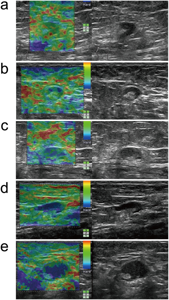
Figure 2.

Gray-scale-RTE dual-mode ultrasonogram of Pattern I ALNs. Images present the general appearance of lesions for RTE scores of (a) 1, (b) 2, (c) 3, (d) 4, and (e) 5 of the lymph nodes with hila: (a) Green portion occupying almost all of the cortex; (b) Blue portion occupying less than 50% of the cortex; (c) Blue portion occupying more than 50% of the cortex, with a scattered green/red portion; (d) Blue portion occupying almost all of the cortex; (e) Blue portion occupying almost all of the cortex, with a green/red ring on the edge of the node.
