
Figure 5.
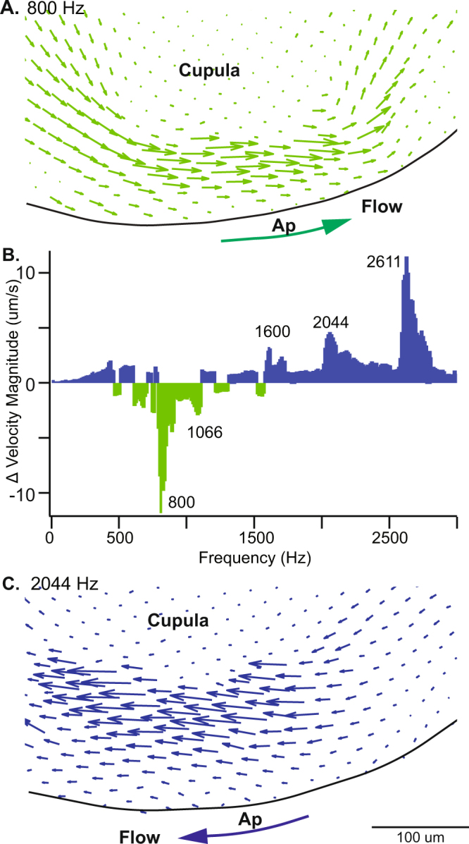
Sustained endolymph pumping in response to auditory frequency stimulation. Particle imaging velocimetry shows sustained endolymph pumping at the apex (Ap) of the lateral canal ampulla flowing over the detached cupula in an animal model of canal dehiscence. (A,C) Vector fluid velocity fields at the at a peak inhibitory frequency 800 Hz and peak excitatory frequency 2044 Hz show endolymph pumping changes direction. Large arrows indicate direction of net endolymph flow. (B) The change in velocity magnitude (µm/s) determined by PIV at the specified frequencies.