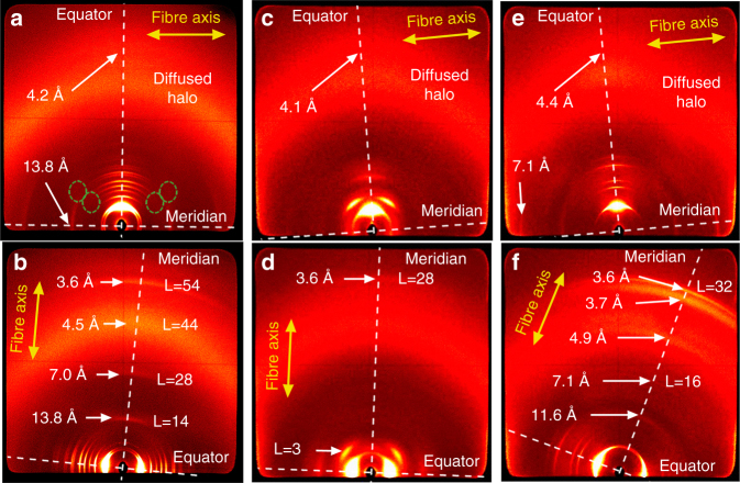
Fig. 3.
WAXS patterns of PBIs 2, 3, and 4. WAXS diffraction pattern of aligned fibers of a,b PBI 2 at 160 °C, c,d PBI 3 at 180 °C, and e,f PBI 4 at 224 °C. The relative position of the fibers are indicated by yellow arrows and the meridian and equator by white dashed lines. The reflections on the equator are indexed according to Colh or Colr phases in Fig. 2. The reflections on the meridian that are attributed to layer lines are indicated as L = X. Dashed green lines indicate additional diffuse reflections at small angles

