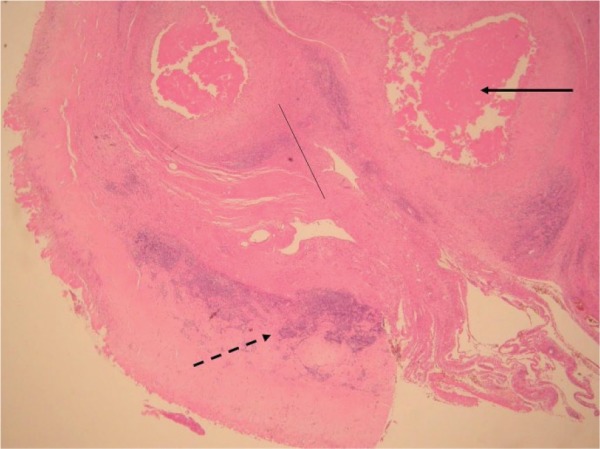
Fig. 3.

Histology showing grade 3 (severe) aseptic lymphocyte-dominated vasculitis-associated lesion. There are coalescing perivascular lymphocytic aggregates and development of lymphoid follicles (broken arrow). There is extensive surface necrosis (unbroken arrow) and sheets of macrophages containing metal debris (thin, unbroken line).
