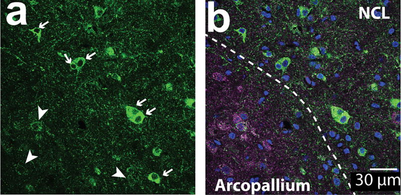
Figure 1. Aromatase+ cells included in analysis along with excluded “ghost cells”.
60× single z-image at the border between arcopallium and NCL from tissue stained against PSD-95 (magenta), aromatase (green), and DAPI (blue). (a) High-intensity fluorescent cells (arrows, “aromatase+ cells”) are located in the NCL, while low-intensity fluorescent cells (arrow heads, “ghost cells”) are shown here in the same section image overlapping with PSD-95 signals (b) and located primarily in the arcopallium. Ghost cells are likely to reflect inputs consisting of pre-synaptic aromatase-positive terminals, as described by (Saldanha et al., 2000).

