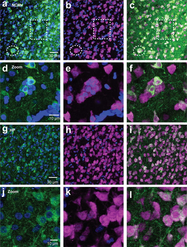
Figure 6. Aromatase cells are found in neuronal clusters of NCM and not HP.
Maximum intensity projection images of 60× z-stack images taken from NeuN (magenta) and aromatase (green) double-labeled sections of NCMv (a–f) and HP (g–l). Exemplars of NeuN clusters in NCMv are noted with dotted lines (a–c). Images in the second row of each region (NCMv: d–f, HP: j–l) are zoomed-in images of the cluster in the dotted square in the first row for NCMv and illustrating the lack of compact clustering for HP.

