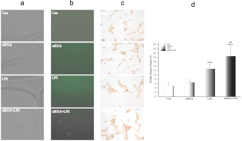
Fig. 6.
Apoptotic cells in HK-2 after transfection observed by TUNEL. a HK-2 observed under light microscopy (200×). b HK-2 observed under a fluorescent microscope, apoptotic cells are fluorescent green (200×). c TUNEL-positive cells observed under light microscopy, and apoptotic cells are dark brown (200×). d TUNEL-positive cells counted and expressed as means ± SD. TUNEL: terminal-deoxynucleoitidyl transferase mediated nick end labeling. ***p < 0.001, relative to the control group. ##p < 0.01, relative to the LPS group

