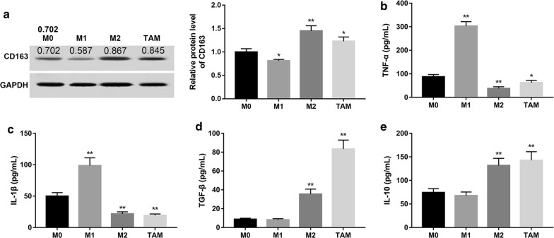
Fig. 1.
Characterization of HCC-conditioned TAMs. Human monocytes were isolated from normal donor buffy coat using anti-CD14 magnetic beads. Monocytes were cultured in the presence of M-CSF for 7 days. TAMs, M1 and M2 macrophages were differentiated as described in “Methods”. a The protein expression of CD163 in each group was examined by Western blot. Densitometric quantification was shown. GAPDH served as the loading control. Levels of cytokines including TNF-α (b), IL-1β (c), TGF-β (d) and IL-10 (e) secreted in culture medium were measured using the specific ELISA system kits. *P < 0.05 and **P < 0.01 vs. the M0 group

