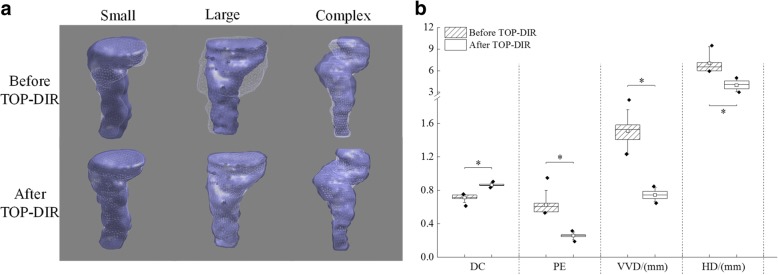
Fig. 2.
a Three example rectum TOP-DIRs with small, large and complex deformation. b Boxplots of DC, PE, VVD and HD over the patient groups before and after TOP-DIR. The boxes run from the 25th to 75th percentile; the two ends of the whiskers represent the 10 and 90% percentiles, the horizontal line and the square in the box represent the median and mean values, respectively. The diamonds represent outliers. Significant differences are marked with “*”

