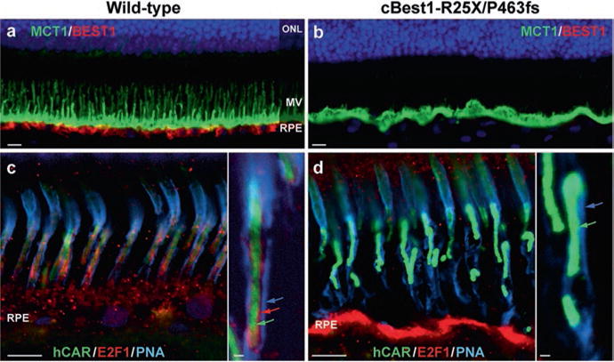
Fig. 38.1.

Abnormal RPE-photoreceptor interface in canine bestrophinopathies (a, b) Representative images of anti-MCT1 (monocarboxylate transporter 1) and anti-Best1 double- labeling in 2-yr-old cBest1-R25X/P463fs-affected retina (b) vs age-matched wild-type control (a). Note the absence of Best1 basolateral labeling and lack of MCT1-positive apical RPE microvillous (MV) extensions in the mutant retina (b). (c, d) Direct comparison of multilabeled (hCAR human cone arrestin, E2F1 transcription factor 1, PNA peanut agglutinin lectin) confocal photomicrographs showing the absence of E2F1-positive cone-associated RPE microvilli (d) when compared to the wild-type control (c, inset, red arrow) and compromised insoluble interphotoreceptor matrix domain (insets, blue arrow) in the mutant retina (d). Cone outer segments (insets, green arrows) appeared disordered (d) in comparison to the control tissue (c). Scale bars: 10 μm (a–d) and 1 μm (insets)
