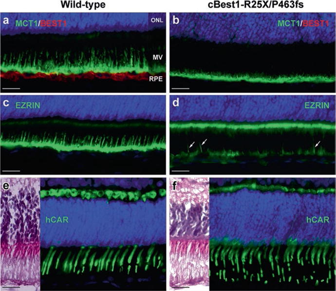
Fig. 38.2.

Underdeveloped RPE apical domain promotes formation of subretinal lesions in bestrophinopathies. Immunohistochemical evaluation of 6-week-old wild-type (a, c, e) and cBest1-R25X/P463fs-affected (b, d, f) retinae revealed underdevelopment of RPE apical domain of cBest-mutant retina, demonstrated by anti-MCT1 (a, b), and anti-Ezrin (c, d) labeling. Note the absence of Best1 basolateral labeling (b), reduced Ezrin immunostaining at the RPE apical surface with only a few Ezrin-positive RPE processes (d, arrows), and a increase of Ezrin labeling associated with microvilli of Müller cells (d). Cone outer segments (green) appear compromised (f, hCAR) when compared to the wild-type retina (e). Scale bar: 20 μm applies to all panels
