
Figure 4:
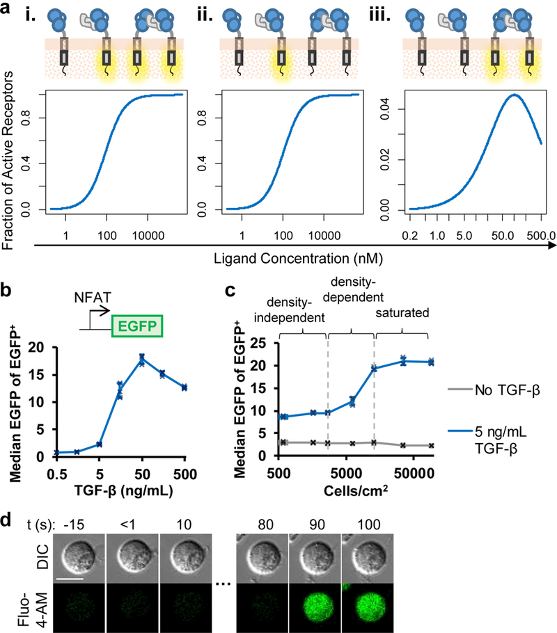
TGF-β–mediated CAR dimerization in cis can activate the TGF-β CAR. (a) Biochemical modeling predicts different dose-response behaviors for receptor activation depending on which receptor states are considered active: (i) all ligand-bound receptors, (ii) only single-receptor/ligand complexes, (iii) only ligand-dimerized receptors. See Online Methods for model parameters. (b) TGF-β dose response of an EGFP NFAT signaling reporter in a TGF-β CAR Jurkat cell line. (c) TGF-β CAR Jurkat cells expressing the NFAT-responsive EGFP reporter were seeded at varying cell densities (Supplementary Fig. 6c) in the presence or absence of soluble TGF-β. (d) TGF-β CAR Jurkat cells loaded with the Fluo-4-AM Ca2+ indicator were imaged every 10 s. After TGF-β addition at t = 0 s to the edge of the cell culture well, individual cells demonstrated Ca2+ influx in the absence of any cell-to-cell contact. All images are representative of ≥5 cells across 3 independent cell cultures. Scale bar denotes 10 μm. In all graphs, data points from n = 3 biologically independent cell cultures are shown with means ± 1 SD.