
Figure 5:
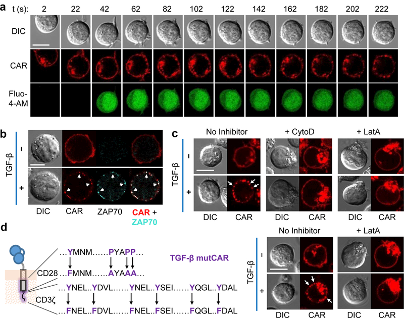
TGF-β CARs form actin-dependent clusters. (a) Time-lapse confocal microscopy of Jurkats loaded with the Fluo-4-AM Ca2+ indicator and expressing a TGF-β CAR-mCherry fusion. TGF-β was added at t = 0 s to the corner of the well and cells were followed at 20 s intervals. (b) TGF-β CAR-mCherry Jurkat cells were incubated with or without TGF-β for 15 min, fixed, and intracellularly stained for ZAP70. White arrows indicate sites where CAR and ZAP70 clusters spatially overlap. (c) TGF-β CAR-mCherry Jurkat cells were imaged 15 min after the addition of TGF-β and/or the actin-polymerization inhibitors cytochalasin D (CytoD) or latrunculin A (LatA). (d) Microscopy as in (c), but with Jurkat cells expressing the non-signaling TGF-β mutant CAR (mutCAR) with the indicated mutations. Images shown are representative of ≥5 cells across 2 independent cell cultures for the CytoD case in (c) and across 3 independent cell cultures for all other images. All scale bars denote 10 μm.