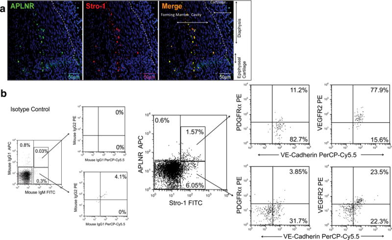
Figure 1. APLNR+, Stro-1+ population contains cells with a phenotype consistent with that of hematovascular mesodermal precursors and/or hemogenic endothelial precursors.

(a) APLNR+ Stro-1+ cells can be identified at 10±1.5gw in the inner part of the developing bone marrow (n=3). DAPI (blue) labels all nuclei. (b) Representative dot plots of flow cytometric evaluation of freshly isolated fetal BMMNC, demonstrating that APLNR+ cells are present, and that the majority of these cells express Stro-1 (n=3). Stro-1+ APLNR+ cells are VE-Cadherin+, express VEGFR2, and a small percentage also express PDGFRα. By contrast, Stro-1+ APLNR- cells contain a mixture of VE-Cadherin positive and negative cells, and only a small fraction express VEGFR2. Percentages depicted in the images are before subtraction of respective isotype controls (n=3). Percentage of background fluorescence obtained when staining the same population of cells with appropriate isotype control is shown in the leftmost panel.
