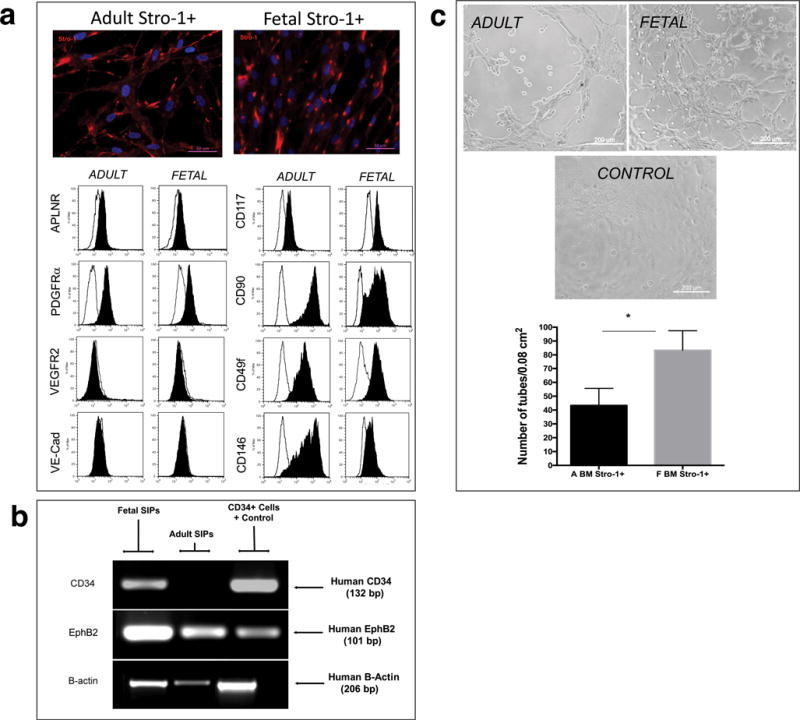
Figure 4. Comparison of cultured fetal and adult SIPs.

(a) Immunofluorescence and Flow cytometric analysis of Stro-1+ cultured fetal and adult cells at P4 are depicted (n=3). The black filled histogram shows fluorescent data for each specific marker, and the unfilled line marks respective isotype controls. Cell populations were considered positive if the ratio between the Median Fluorescence Intensity (MFI) for the specific marker and the MFI of the isotype control ≥ 1.5. (b) RT-PCR analysis, using human-specific primers for CD34, demonstrated that fetal, but not adult, SIPs expressed CD34. Amplification of the same samples with primers specific for human EphB2 and B-actin confirmed the presence amplifiable RNA. For each set of primers samples were run simultaneously in the same gel, but gel was cropped to improve clarity, and to show amplification of samples specific to fetal and adult BM-derived SIPs. (c) Tube formation assays confirmed that, at the same passage (P4), both cell populations were able to form capillary tubes. Cells initiated tubule formation at 2–4h of culture, which fully developed by 6h, with the presence of nodes of ≥ 4 branches; images were acquired on a Ziess Axiovert 200M and 10X magnification. Quantification of number of branch sites/nodes demonstrated fetal cells were significantly more efficient at generating tubes than their adult counterparts (n=3).
