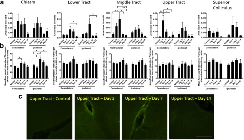
Fig. 11.
Fibrinogen immunoreactivity in the brain following partial optic nerve transection. Mean ± SEM fibrinogen immunoreactivity assessing a area and b intensity above a constant set threshold in control uninjured optic nerve, compared to 1, 3, 7, 14, and 28 days, assessed separately for the right- and left-hand sides of the brain. Lower, middle, and upper regions of the optic tract were assessed separately. n = 5–6/group. *p ≤ 0.05, **p ≤ 0.01, ***p ≤ 0.001, ****p ≤ 0.0001. Representative images of the upper tract (c) show breaches in the BBB indicated by increased areas of diffuse fibrinogen immunoreactivity (bracketed), 3 days after injury, compared to control uninjured, and 7 and 14 days following partial optic nerve transection. Scale bar for c: 100 μm

