Figure 13.

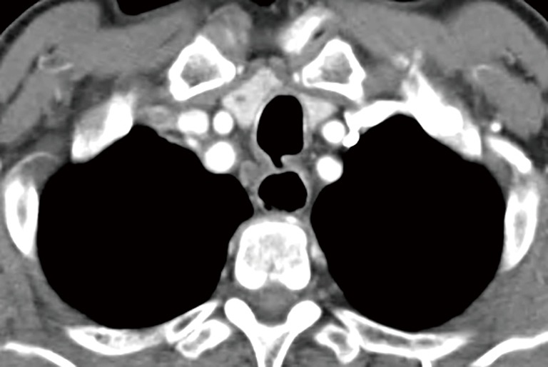
Because the TEF is always more cephalad on the tracheal side, a single axial view on the CT scan may not show up the entire TEF and could be misinterpreted as a tracheal diverticulum. TEF, tracheoesophageal fistula.

Because the TEF is always more cephalad on the tracheal side, a single axial view on the CT scan may not show up the entire TEF and could be misinterpreted as a tracheal diverticulum. TEF, tracheoesophageal fistula.