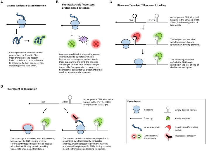
FIGURE 3.
Methods that visualize translation in vivo. (A) Gaussia luciferase fused to the protein of interest enables live visualization of translation. (B) Photoswitchable fluorescent proteins allow for live visualization of translation. (C) Ribosome “knock-off” uses fluorescent tracking to visualize the progress of the ribosome. (D) Fluorescent co-localization uses fluorescent proteins to mark the transcript of interest and the ribosome or the nascent peptide.

