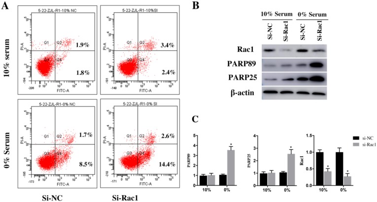
Figure 3.
Rac1 inhibited apoptosis of H9c2 cells induced by serum deficiency. A. Flow cytometry was used to analyze the apoptosis of H9c2 cells cultured in 10% and 0% serum conditions for 48h after transfection with si-Rac1 for 48h. Q-2 presents the percentage of late apoptosis cells, Q-4 presents the percentage of early apoptosis cells and Q-3 presents live cells. B. Western Bolt results of inhibitory effects of si-Rac1 on Rac1 expression after transfection for 48h and the changes of the cleaved PARP25 and PARP89 in si-NC and si-Rac1 group in 10% and 0% serum. C. Diagram of inhibitory effects of si-Rac1 and the cleaved PARP25 and PARP89. Densities were first standardized to β-actin and then to si-NC group. N=3. *P < 0.05 vs si-NC group.

