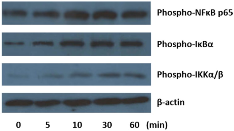
Figure 3.
Detection of expression level of p-NFκB p65, p-IκBα and p-IKKα/β by Western blot analysis. U937-differentiated macrophages were exposed to silica and incubated for 5, 10, 30 and 60 min, respectively, before total protein was harvested. Then, the protein expression level of phosphorylated form of NFκB p65, IκBα and IKKα/β was detected by primary antibodies and respective secondary antibodies followed by enhanced chemiluminescence detection and image captured by X-ray film. Total protein amount of each sample was normalized by expression of β-actin. The image was a representative of three independent trials.

