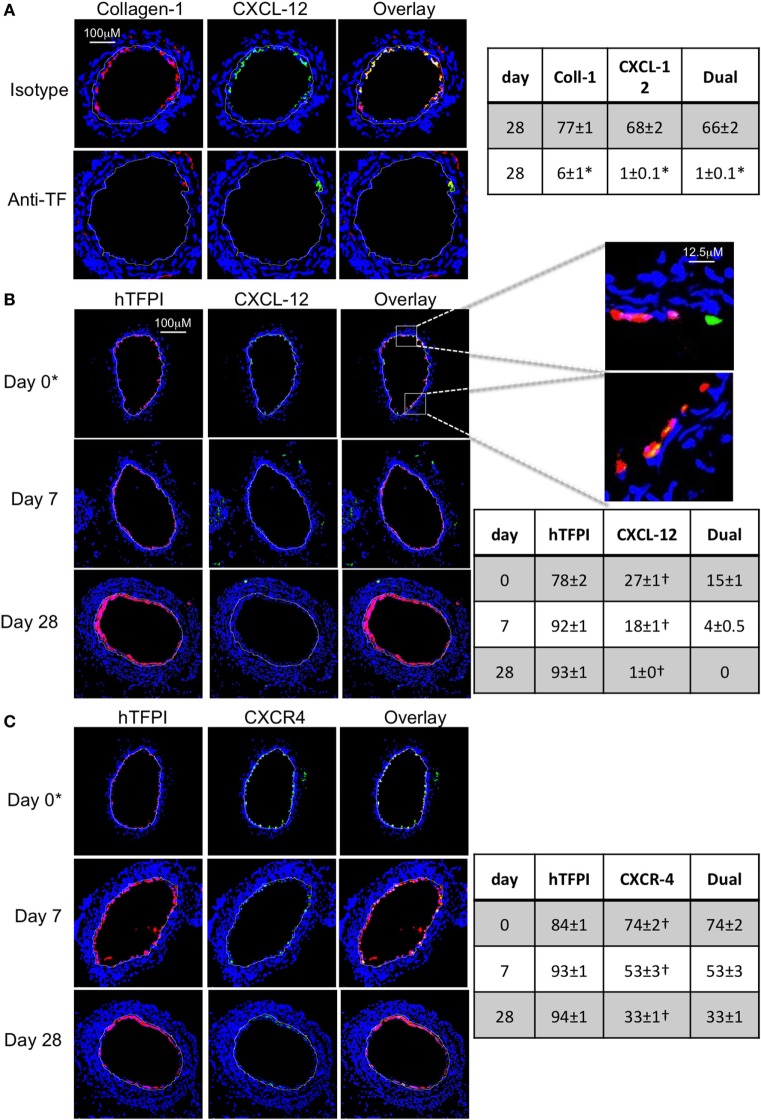
Figure 10.
Recruitment after adoptive transfer with inhibition of tissue factor (TF). Panels show immunohistology of representative sections through injured mouse carotid arteries harvested on days post-injury as indicated. Sections from same day are consecutive. All sections stained with DAPI (4,6 diamidino-2-phenylindole) nuclear stain (blue) and (red) anti-collagen-1 (A) or human tissue factor pathway inhibitor (hTFPI) (B,C), and (green) anti-CXCL-12 (A,B) or CXCR4 (C). Yellow indicates co-localization. The annotated white line defines the junction between neointima and media. (A) Day 28 sections, in wild-type (WT) mice adoptively transferred CD34+ cells pre-incubated with istotype control or anti-TF antibody. (B,C) Adoptive transfer of purified CD31+ myofibrocytes from CD31-TFI-Tg mice to WT mice immediately post-injury. *Sections harvested within an hour of injury. Expanded areas illustrate that some of the early luminal CXCL-12 staining is independent of recruited hTFPI+ cells. Table besides (A) describes summary of staining from all mice (n = 6), showing the proportion of the neointimal area that is positive for collagen-1, CXCL-12, or both on day 28 (% ± SEM), whereas those besides (B,C) described proportion that is positive for hTFPI, CXCL-12 (B), CXCR4 (C), or dual positive on days 0, 7, and 28. Data derived from three random sections from each of the arteries from each mouse (see Materials and Methods). *cf isotype for anti-TF p < 0.001. †cf adoptive transfer of WT cells (Figures 9B,C) at equivalent times p < 0.001. Experiments repeated at least twice.

