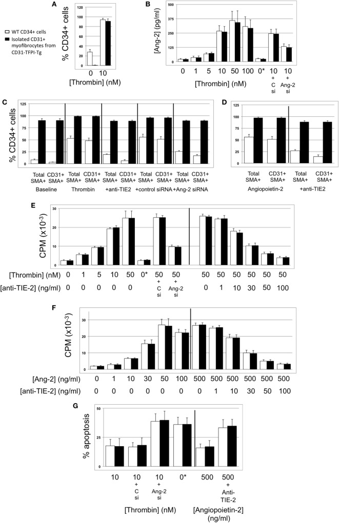
Figure 7.
Thrombin-induced proliferation and survival are angiopoietin-2-dependent. In all, responses of wild-type (WT) CD34+ cells are shown as white bars, whereas purified CD31+ myofibrocytes from CD31-TFPI-Tg mice are shown as black bars. 0*Indicates incubation with active site inhibited thrombin. Abbreviations: Csi, control scrambled siRNA; Ang-2si, siRNA specific for angiopoietin-2. (A) Expression of angiopoietin-2 at baseline and after incubation with thrombin for 5 days, expressed as the percentage of CD34+cells staining positive in immunocytofluoresence analysis. (B) Secretion of angiopoietin-2 into supernatant after 5-day incubation with thrombin at the indicated concentrations, analyzed by ELISA. (C) Expression of smooth muscle actin (SMA) and joint expression of CD31 with SMA at baseline and after incubation with thombin for 5 days, either alone or with anti-TIE-2 antibody or siRNA against angiopoietin-2 or control. Data expressed as the percentage of CD34+ cells staining positive in immunocytofluoresence analysis. (D) As (C) but cells incubated with angiopoietin-2 ± anti-TIE-2 antibody. (E) Proliferation, assessed by 3H-thymidine incorporation and expressed as counts per minute (CPM) after incubation with thrombin at the indicated concentrations. (F) Proliferation, assessed by 3H-thymidine incorporation and expressed as CPM after incubation with angiopoietin-2 at the indicated concentrations. (G) Degree of apoptosis after incubation with thrombin or angiopoietin-2 at the indicated concentrations. Anti-TIE-2 antibody used at 50 ng/ml. All experiments repeated at least twice.

