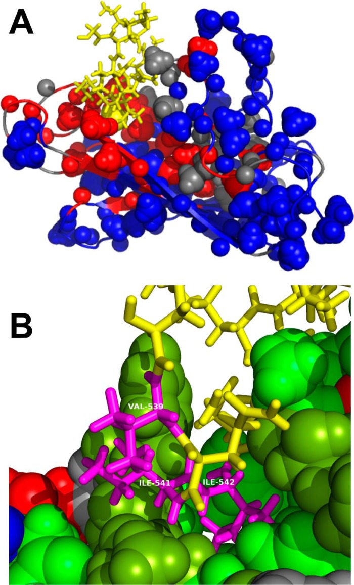
Figure 9.
A, molecular replacement structure for Hsc70 BETA domain with the TAU peptide bound. Color coding is as in Fig. 6. The sequence GKVQIINKKG is shown in yellow. The sphere is the CD methyl group of the second Ile of the in cis sequence GKVQIINKKG. B, details of the molecular replacement structure for Hsc70 BETA domain with the in cis TAU1 sequence bound. On Hsc70, hydrophobic residues are in green, amphipathic residues (Thr, Tyr) in peat green, positive residues in blue, negative residues in red, and polar residues in gray. Residues VII of the in cis sequence GKVQIINKKG are labeled as Val539, Ile541, and Ile542 (magenta). Other residues of TAU1 are in yellow.

